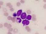
Cancer immunotherapy is the use of the immune system to reject cancer. Gammadelta (γδ) T cells are an immune cell found within epithelial tissues. Epithelial tissue is found throughout the body. It is present in the skin, as well as the covering and lining of organs and internal passageways, such as the gastrointestinal tract. Epithelial tissues are also a prominent component of glands, such as prostate gland. γδ T cells play unique and critical roles in recognition of damage or disease in epithelial tissues and provide a crucial first line of defense.
 Unlike the alphabeta (αβ) T cells of the immune system (that are launched on a search-and-destroy mission when the skin suffers a cut or other damage), most γδ T cells do not circulate through the bloodstream. γδ T cells comprise 2–5% of T cells in human peripheral blood. Instead, they are a major T cell component of the skin, lung, and intestine, where they take up residence and monitor the neighboring epithelial cells for damage and disease. γδ T cells have both ‘innate’ and ‘adaptive’ characteristics in the immune response (commonly considered to bridge innate and adaptive immunity), and their activities are mediated by multiple pathways that are under elaborate regulation by other immune components. γδ T cells provide protective innate immunosurveillance against certain malignancies, particularly those of epithelial origin, and kill a range of tumor cells in a manner that does not require the recognition of tumor-specific antigens.
In contrast to conventional αβ T cells, γδ T cells do not recognize antigenic peptides. When there’s damage or disease, the cell signaling receptors of the immune system are up-regulated, and signals that are transmitted through their interaction with each other costimulate a T cell response that aids in tissue repair or killing of tumors. In fact, some γδ T-cell receptors (TCRs) such as Vγ9Vδ2 act like pattern recognition receptors, hence detecting pyrophosphates derived from multiple microbes (and tumor cells) as molecular patterns. Vγ9Vδ2 T cells recognize in a wide array of transformed cells and are activated in various tumors. They can directly kill their targets and release pro-inflammatory cytokines that boost the anti-tumor effector cells of the adaptive immune system.
Gammadelta T cells: innately adaptive immune cells?
Understanding the complexity of γδ T-cell subsets in mouse and human.
The molecular interaction of CAR and JAML recruits the central cell signal transducer PI3K.
The multifunctionality of human Vγ9Vδ2 γδ T cells: clonal plasticity or distinct subsets?
However, γδ T cells play a complex role in cancer and can promote, as well as inhibit, tumor growth. In addition to tumor cell killing, γδ T cells express a number of cytokines and other soluble factors in response to tumors. Soluble factors expressed by γδ T cells in these settings include IFN-γ, TNF-α, IL-4, IL-10, TGF-β, IL-17, and a number of growth factors. These factors have differing and sometimes opposing effects on antitumor immunity and tumor angiogenesis, and likely contribute to the complex role of these cells in cancer. Moreover, in cancer patients with a growing tumor, immune tolerance between the host immune system and tumor may have already been established. Thus the activation of innate immune system acts like as a ‘reset button’ in cancer patients to restart the immune responses.
Role of gamma-delta T-cells in cancer: another opening door to immunotherapy.
Harnessing γδ T cells in anticancer immunotherapy.
Complex role of γδ T-cell-derived cytokines and growth factors in cancer.
What lessons can be learned from γδ T cell-based cancer immunotherapy trials?
Human Vδ2 versus non-Vδ2 γδ T cells in antitumor immunity.
γδ T-cell immunotherapy for lung cancer.
How tumors might withstand γδ T-cell attack.
Toll-like receptors (TLRs) play a fundamental role in activation of innate immunity and there are 11 known functional TLRs in humans (TLR1-11). TLR1, 2, 4, 5, and 6 are primarily expressed on the plasma membrane of immune cells. These TLRs recognize a variety of unique microbial membrane components like lipids, lipoproteins and proteins. Conversely, TLR 3, 7, 8 and 9 are expressed on intracellular vesicular membranes and are commonly involved in recognition of viral components and nucleic acids. And the respective ligands can directly modulate their effector functions. TLR ligands enhance cytotoxic tumor responses of γδ T cells and regulate the suppressive capacity of γδ T cells. Although some types of TLRs are also expressed in cancer cells. TLRs on different cancer types could promote tumor growth, but their role remains poorly understood. Thus, TLR ligands have become attractive targets for cancer immunotherapy.
γδ regulatory T (Treg) cells have recently been identified in human diseases including cancer. Tumor-derived γδ Treg cells induce immunosenescence in the targeted naive and effector T cells, as well as dendritic cells (DCs). Furthermore, senescent T cells and DCs induced by γδ Treg cells have altered phenotypes and impaired functions and develop immune-suppressive activities, further amplifying the immunosuppression mediated by γδ Treg cells. But manipulation of TLR 8 signaling in γδ Treg cells can block γδ Treg-induced conversion of T cells and DCs into senescent cells.
Modulation of γδ T cell responses by TLR ligands.
TLR agonists: our best frenemy in cancer immunotherapy.
Toll-like receptor expression and function in subsets of human gammadelta T lymphocytes.
Toll-like receptors in prostate infection and cancer between bench and bedside.
Characterization of the Toll-like receptor expression profile in human multiple myeloma cells.
Human regulatory T cells induce T-lymphocyte senescence.
TLR8: the forgotten relative revindicated.
γδ T cells were found to express TLR1, 2, 3, 5, 6 and 7. Interestingly despite TLR3 being expressed in both conventional αβ T cells and γδ T cells only the latter have been shown to respond to polyinosinic-polycytidylic acid, a synthetic analogue of double stranded ribonucleic acids, and a TLR3 agonist. TLR3 plays a fundamental role in pathogen recognition and activation of innate immunity and thus involved in the innate immune response against various viruses.
TLR3 can also be detected in many tissues. At the cellular level, myeloid dendritic cells and macrophages have been found to express TLR3. TLR3 is also present in non-immune cells such as epithelial cells of various origins (lung, intestine, breast, kidney, prostate and pancreas) but also in mesenchymal cells and in endothelial cells. Of interest, TLR3 is the TLR that is expressed most strongly in the brain, and the activation of TLR3 exerts an antitumoral effect. Here is the good news. TLR3-mediated signaling pathways can be modulated by several natural substances.
Direct costimulatory effect of TLR3 ligand poly(I:C) on human gamma delta T lymphocytes.
Differential effects of phenethyl isothiocyanate and D,L-sulforaphane on TLR3 signaling.
TLR3 ligand induces a direct costimulatory effect on phosphoantigen stimulated γδ T cells, but not on aminobisphosphonate-stimulated γδ T cells. TLR3 ligands could stimulate their receptors both in cancer cells and in immune cells inhibiting tumour growth both directly and through the immune system (direct and immune-mediated cell death). Bisphosphonates are a class of drugs used to treat osteoporosis, which bind avidly to hydroxyapatite bone mineral surfaces and their major action is to inhibit osteoclast activity and thus bone resorption.
Amino-bisphosphonates (or nitrogen-containing bisphosphonates) are newer generation bisphosphonates. Amino-bisphosphonates like zoledronic acid (Zometa) are known to stimulate γδ T cells and exert a variety of direct and indirect anticancer activities. But, the old bisphosphonate drugs like clodronate (Bonefos) have no ability to kill cancer cells. One of the problems you have is that most cancer cells start becoming resistant to amino-bisphosphonates: they adapt. Over time, they can become more resistant to many different types of cancer treatments.
Aminobisphosphonates as new weapons for gammadelta T Cell-based immunotherapy of cancer.
γδ T cells comprise 2–5% of T cells in human peripheral blood. They are expanded in the peripheral blood of patients with a variety of bacterial, viral, and parasitic illnesses to represent 50–60% of peripheral blood T cells. γδ T cell numbers, activity and functions can also be modified and enhanced by natural substances, such as plant-derived tannins, dietary nucleotides, fatty acids, and dietary alkylamines including L-theanine, a distinctive amino acid that is found in green tea. Particulary, L-theanine enhances γδ T cell proliferation, and interferon-gamma secretion. L-theanine also primes γδ T cells so they can immediately respond if confronted with a bacterial, fungal or viral infection. γδ T cells, once activated or primed can further activate normal αβ T cells, CD4 and CD8 T cells which produce antibody and attack cancer and virally infected cells.
The intracellular cholesterol levels and lipid metabolism also afect T cell receptor (TCR) signaling, and activation and proliferation of γδ T cells. Valproic acid, a commonly prescribed antiepileptic agent with histone deacetylase (HDAC) inhibitory activity, also enhances the antitumour effect of γδ T-cells via up-regulation of NKG2D (natural killer group 2D) ligands.
In summary, specific cellular immunotherapy of cancer requires efficient generation and expansion of cytotoxic T lymphocytes that recognize tumor-associated self-antigens. Dietary modifiers-activated γδ T cells permit generation of cytotoxic T lymphocytes specific for weakly immunogenic tumor-associated epitopes. Hepazym contains a proprietary blend of dietary modifiers of γδ T cell activity and induces immune responses to tumor-associated self antigens. Clinically, Hepazym immunotherapy may be most effective when combined with Herbalzym therapy as targeted natural anticancer therapy to treat a large variety of cancers including lung, colon, prostate, breast, brain, skin, liver, pancreatic, ovarian and cervical cancer.
Professional antigen-presentation function by human gammadelta T Cells.
γδ T-APCs: a novel tool for immunotherapy?